Mammography
Breast Cancer Screenings In Salida
Mammograms are one of the most effective tools available to diagnose breast cancer. While a mammogram cannot prevent cancer, it can spot early indicators of the disease, allowing doctors to start early treatment that will hopefully stop it from spreading. You should ask a doctor familiar with your medical history when and how often you should receive mammograms.
Mammograms are just one of the imaging tests performed at Heart of the Rockies Regional Medical Center. If you would like to schedule an appointment or receive other tests, call (719) 530-2396.
3-D Mammography
Heart of the Rockies Regional Medical Center is equipped with 3-D digital mammography, or tomosynthesis, which obtains highly detailed images of breast tissue. This mammography is capable of catching numerous warning signs of cancer. The radiation levels of both 2-D and 3-D mammograms are well below the exposure levels for imaging established by the American College of Radiology.
Tomosynthesis works by taking multiple X-rays of the breast to create a 3-D image. Regular mammograms, on the other hand, only produce a single image, limiting the physician’s ability to diagnose issues. Women with dense breast tissue may be unable to receive accurate results without a 3-D mammogram.
What A Mammogram Looks For
There are a few anomalies that occur in breast tissue that act as cancer warning signs. If you have had mammograms in the past, a doctor can perform a better diagnostic by comparing past results with new ones.
Doctors look for the following in a mammogram:
- Calcifications - small mineral deposits in breast tissue. While large macrocalcifications (larger deposits) are fairly common in women over 50, microcalcifications are more alarming, but do not always indicate cancer.
- Cysts -These small fluid-filled lumps are not cancerous, but can appear as lumps on the breasts much like tumors. Mammograms can help doctors determine if a lump is a tumor or cyst.
- Tumors - While tumors are not always cancerous, it is important to have the lump examined by a doctor as soon as possible.
- Breast density - Denser breasts have a higher risk of developing cancer than others. 3-D mammography has made it easier to examine these tissues and look for warning signs.
If it is discovered that you have dense breast tissue, an automated breast ultrasound (ABUS) may be ordered.
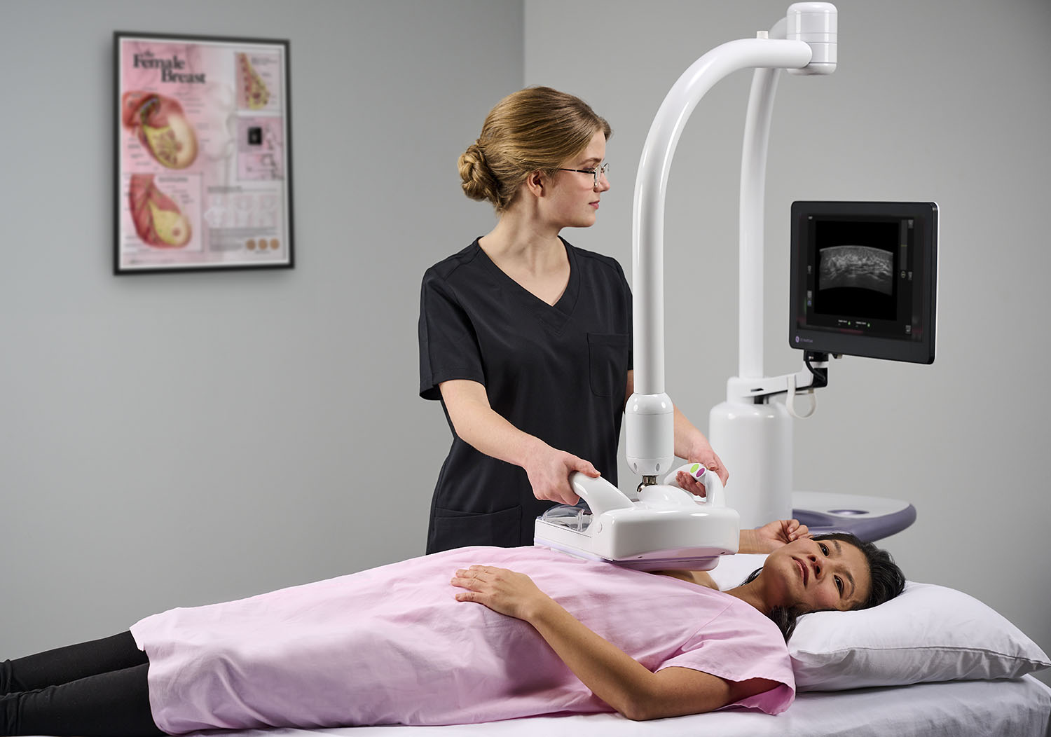
Automated Breast Ultrasounds (ABUS)
Breasts are comprised of a combination of fat and breast tissue. A breast with more tissue than fat can be considered dense. During each mammogram, you will be given information about the density of your breasts, which is determined by the radiologist who reads your mammogram.
There are four categories of breast density, as shown below:
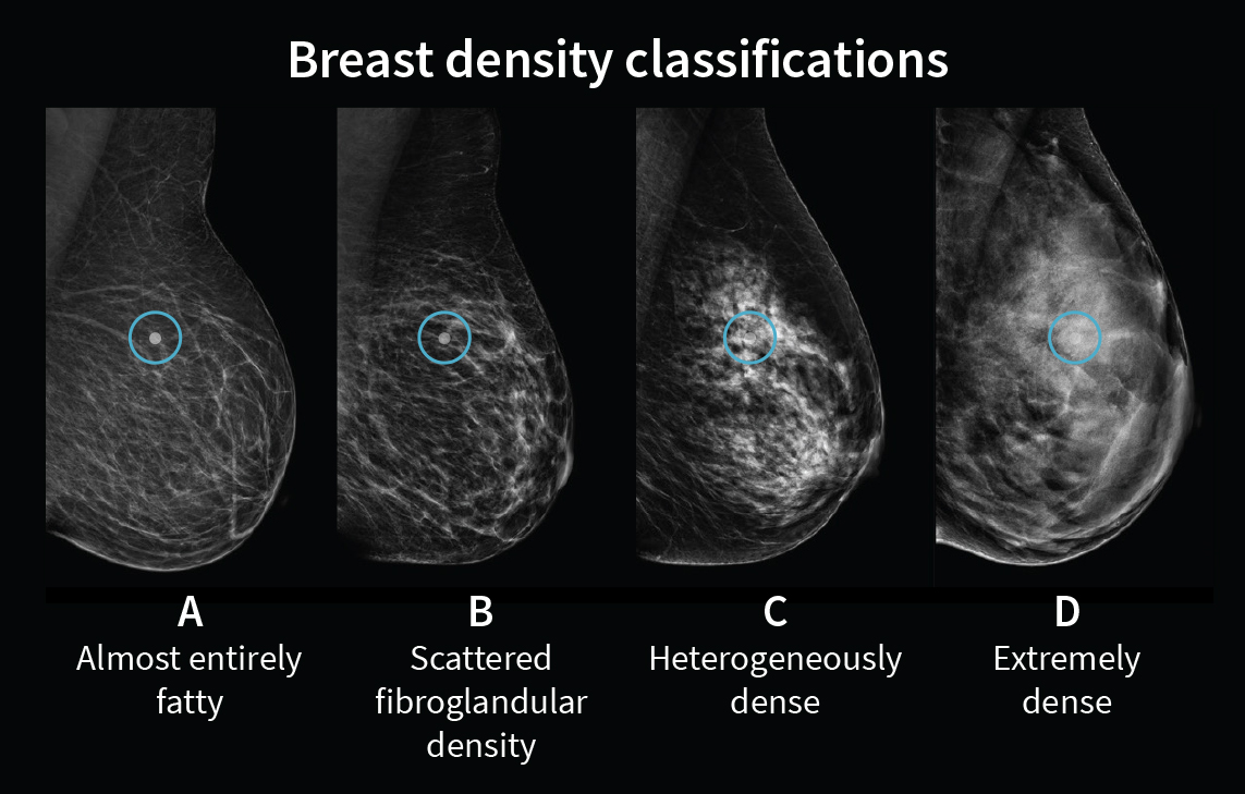
Categories C and D are considered dense. Did you know that more than 40% of women have dense breast tissue? You may be 4-6x more likely of developing breast cancer with dense breast tissue.*
During routine mammograms, cancerous masses appear white. Unfortunately, dense breast tissue also appears white, making it more difficult to identify cancerous lumps and masses as opposed to normal breast tissue.
An ABUS screening is a supplemental exam, and the next critical step in maintaining your breast health. This ultrasound procedure creates a 3D picture of your breast using sound waves, where tissue appears white and masses appear black, making them easier to see and identify. Utilizing both your routine mammogram and ABUS screening results helps provide a more complete evaluation of your dense breast tissue. The ABUS screening has been shown to improve breast cancer detection over mammography alone for women with dense breasts.
Watch this video to learn more about the ABUS screening and what it might mean for you.
Frequently Asked Questions about ABUS screenings
How do I know if I have dense breast tissue?
This is something you learn through your first (and regularly-scheduled) mammogram. Dense breast tissue is determined by categories; categories C and D are considered dense, as in the image further up this page.
How will I know if I need an ABUS screening?
Both you and your primary care provider (PCP) will be notified of your dense breast tissue status via a letter mailed to you, results through your patient portal, or your PCP will call you directly with results. Your PCP will order the ABUS screening as a follow-up service for you.
How do I go about getting the ABUS screening done?
Once it is determined you have dense breast tissue, your ordering provider (typically your PCP) will order your ABUS screening follow-up. Once ordered, we will determine pre-authorization of the screening and our centralized scheduling department will reach out to you to schedule the screening.
Can I refer myself for an ABUS screening?
Unfortunately, no. This needs to be referred through your PCP, or provider who ordered your mammogram.
I already know I have dense breast tissue. Can the ABUS screening take the place of my annual mammogram?
No. It is still crucial you receive your annual mammogram, and the ABUS screening is in addition to that.
Does my insurance provider cover my ABUS screening?
Unfortunately, we do not know that information, as every insurance carrier varies in their coverage. Prior to receiving an ABUS screening, you should call your insurance provider to confirm coverage.
How will I receive results from my ABUS screening?
You should also receive these through your referring provider (i.e., PCP) and receive a letter in the mail.
Additional Resources about ABUS screening
Patient brochure - different tests for different breasts
*Source: Breast Imaging and Reporting and Data System (BI-RADS®), American College of Radiology.